

Cardiovascular diseases are the main cause of mortality and disability around the world. However, patients susceptible to the increased risk of developing heart disease can avoid them, in time by changing the lifestyle and taking appropriate medicines. Alas, in most cases of pathologies remain unbearable until the myocardial infarction occurs or stroke.
Therefore, it is important to determine biomarkers that would help identify patients with hidden cardiovascular diseases. People exposed to them, as you know, prone to the occlusion of the vessels (violation of the passability) of the retina of the eye - multilayer nervous tissue with a complex capillary network. Surface and deep vascular plexuses provide oxygen inner and middle parts of the retina, while the outer layer receives oxygen from choriocapillary. With severe vessel occlusion, two inner layers of this sensory membrane are atrophically atrophy, and with retinal microfarcts of the eye, such as paracentral acute median maculopathy, the average layer is selectively affected.
Using the non-invasive imaging technology with submillimeter resolution in vivo and optical coherent tomography, it is possible to identify anomalies on the retina images indicating ischemia: in the acute phase, they manifest themselves as a hyperreflective perivular strip at the level of the inner nuclear mesh-shell layer. Such lesions occur as a result of hypoperfusion or microembolis like an occlusion of the artery and veins of retina, hypertension, Purcher's retinopathy and sickle cell anemia. The visualization of the micrososcience demonstrates the voids of the blood flow signal in the acute phase, which further confirms the ischemic nature listed above.
The authors of the new research - doctors from the University of California in San Diego (USA) - tried to find out whether such eye lesions are common in patients with cardiovascular diseases and is it possible to predict them in this way. The article is published in the magazine EclinicalMedicine.
"Eyes - a window in our health, many diseases can manifest themselves in them. And cardiovascular diseases are no exception, - noted by the research group Mathieu Bachum, - ischemia, that is, a decrease in blood flow, caused by a heart disease, can lead to an insufficient inflow of blood to the eye and bring the intimidation of the retina cells, leaving behind the steady "tags ". We called them ischemic perivascular retina lesions, or Ripls, and sought to determine if they could serve as a biomarker cardiovascular diseases. "
Medics studied records of 13,940 people who passed an oct scanning (optical coherent tomography) of a yellow spot - the place of the greatest visual acuity in the retina - at the University of California in San Diego on various clinical testimony from July 1, 2014 to July 1, 2019 . After analyzing Medkart, researchers have distributed patients into two groups: 84 people got 84 people with documented cardiovascular diseases, in the second - 76 healthy (that is, there were no ischemic heart disease, stroke, heart failure, atrial fibrillation, hypertension, diabetes of both types, chronic obstructive disease of the lungs or pulmonary hypertension). In addition, no one from the participants revealed the accompanying retinal pathologies.
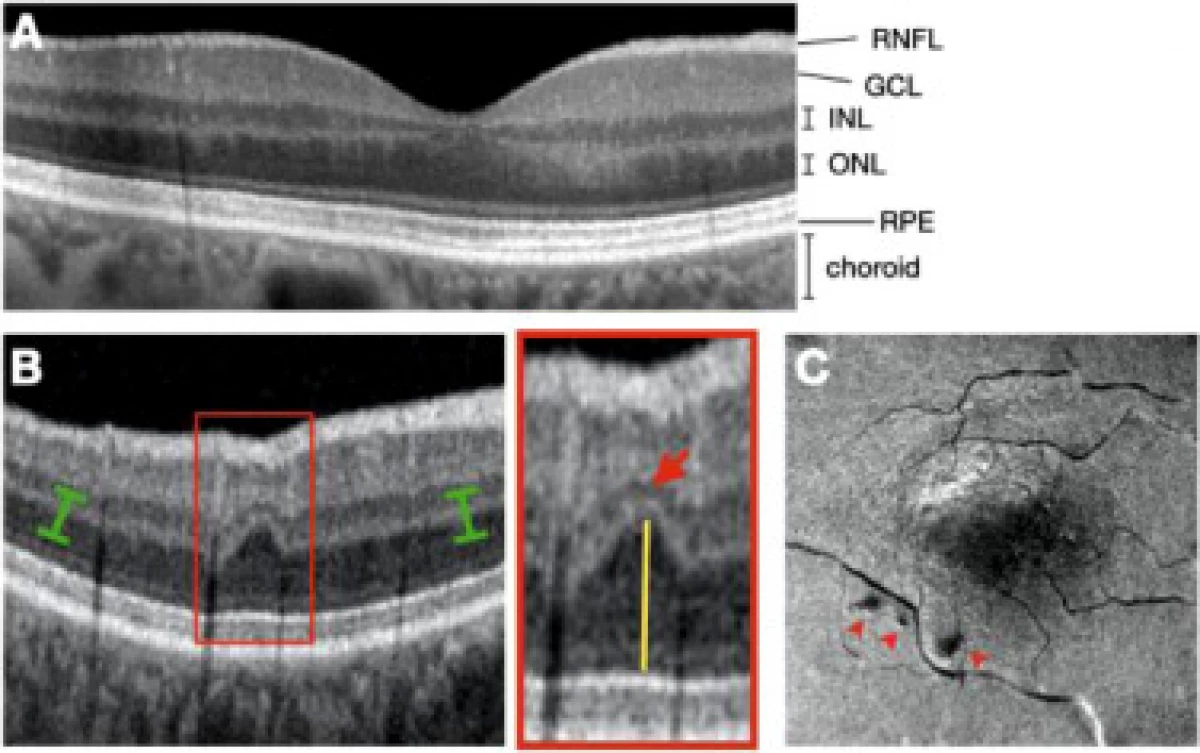
B) SD-OCT B scanning, demonstrating Ripl (red rectangle). Noticeably focal abbreviation inL (red arrow), accompanied by compensating upward expansion of a darker external nuclear layer (yellow line). FROM)
The appearance of the face restored from the three-dimensional bulk scan assembled from consecutive B scanning. Three Ripl look like dark dots (red arrows) / © EclinicalMedicine
It is known that the risk of cardiovascular diseases in the United States is determined using a special ASCVD calculator developed by the American College of Cardiologists. The authors of the study found a correlation between the number of retinal lesions (RIPL) and risk assessment calculated by ASCVD for all volunteers.
"In patients with low and border indicators ASCVD there was a small amount of Ripl in the eyes, but as the risk ASCVD increased, the number of Ripl grew. The total ripl per patient was significantly higher in a group with cardiovascular diseases compared with the control group (2.8 against 0.8). The number of RIPL in individuals with IBS and stroke was 2.4 and 3.7. In patients with myocardial infarction, the Ripl indicator turned out to be 3-4 - compared with 1.3 ripl in patients with IHS without a heart attack. We noticed most of the retinal lesions in patients undergoing stroke. Since the retina is a direct continuation of the brain, it is likely that Ripls talk about cerebral disease than about diseases of coronary vessels, "researchers told.
Thus, the results confirmed that the damage to the retina - biomarkers of the preceding ischemic infarction of this inner shell of the eye are common in patients with cardiovascular diseases and can serve as an additional feature for their prediction and detection. According to the authors of the work, if the ophthalmologist discovers the Ripl patient, then it should be sent to the reception and to the cardiologist.
Source: Naked Science