
The study was supported by the Presidential Program of the Russian Scientific Fund and published in the magnetine of Magnetic Resonance in Medicine. "Globally, our discovery will allow to develop phased lattices for research MRI and accelerate their introduction into clinical practice," explains one of the authors of the article, graduate student of St. George Solomach, - phased lattices allow you to cover a much greater scan area, and also better control the process itself. The interpretation of the final result becomes easier and faster. "
Magnetic resonance topography - the method in modern medicine is as important as expensive. It allows you to explore the internal organs of a person in non-invasive (without direct autopsy) and practically does not have an ionizing effect compared to X-ray tomography. However, one apparatus is worth not less than 15 million rubles (not counting the cost of service) and occupies a place commensurate with a small storage area.
At the same time, the quality and accuracy of images often leave much to be desired. In the tasks of clinical MRI, tomographs with a field level of a half and three Tesla are used. However, for the tasks associated with research where it is required to obtain the maximum resolution, tomographs with a field level seven or more tesla are used.
The principle of operation of MRI is based on the interaction of a radio frequency magnetic field with hydrogen nuclei. At the same time, since the nuclei of hydrogen atoms in our body are small magnets, they are oriented along the field lines turning in one direction.

True, this situation is energetically unprofitable, and atoms are returned to their "usual" state so quickly, as soon as they can highlighting excess energy. It is according to its number that one can understand whether there are many atoms of a certain substance in the right tissue of man. Thus, the activity of the brain is investigated - after all, the more blood (and therefore water with hydrogen atoms) in a certain area, the higher its activity. It is also possible to detect tumors in the early stages, since the affected cells create more fluid than the usual.
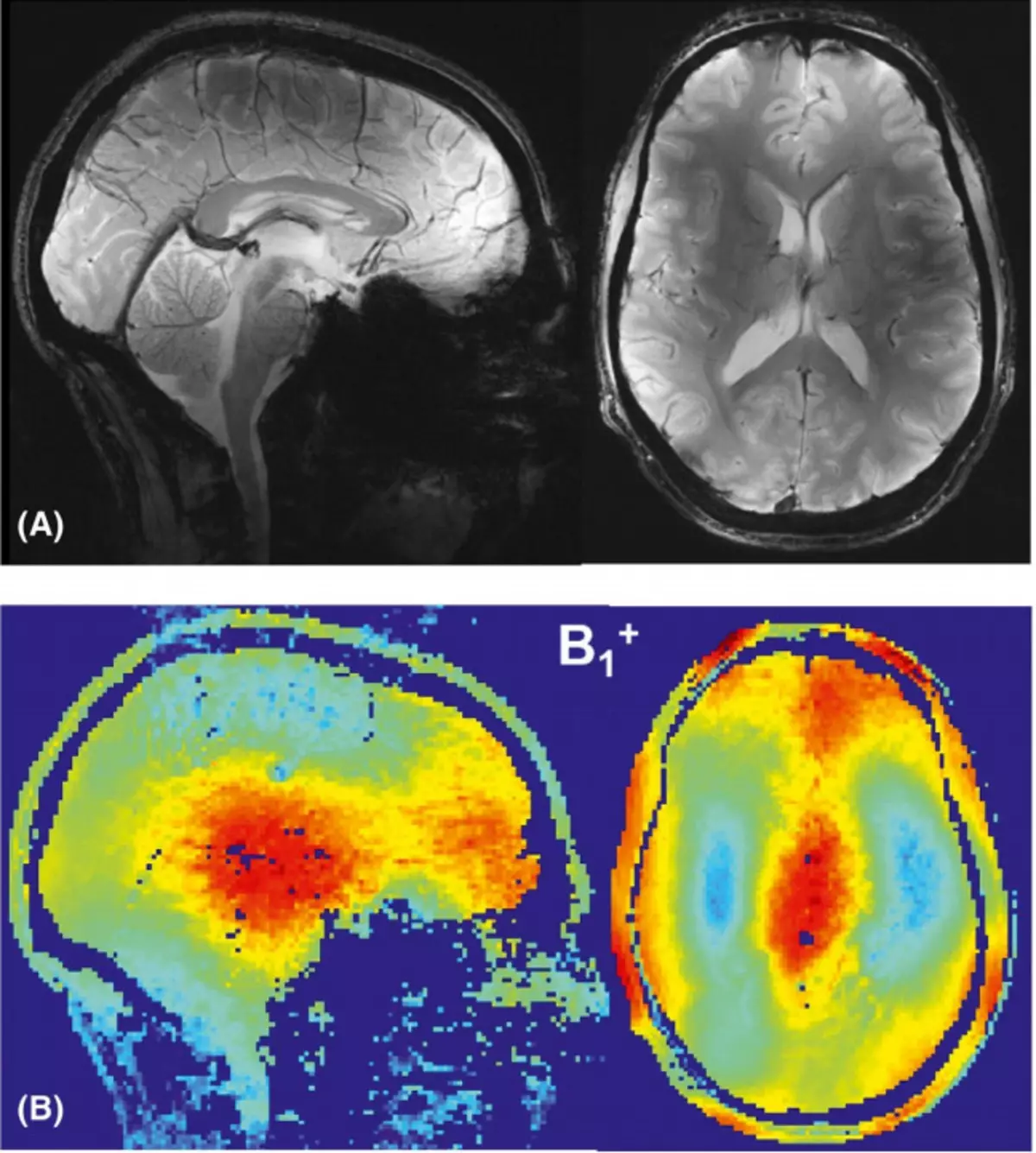
Magnetic fields obtained using the developed phased lattice / © AVDIEVICH ET AL. / Magnetic Resonance in Medicine, 2021
To create a radio frequency magnetic field in tomographs with a field level, more than seven tesla use phased antenna arrays. They have an important advantage: allow you to change the localization of the research subject, without moving at the same time the lattice itself. Dipole antennas can be applied as elements of the lattice. However, there may be a connection between active dipoles, which reduces the efficiency of the entire radio frequency coil.
To prevent this, passive dipoles are used. Usually they are located in parallel active, and it solves the problem. But this method should be used with caution, since too large passive dipoles interact with the field, porting its homogeneity, which ultimately leads to a decrease in the quality of the final picture, which means that the results of the entire medical examination.
Scientists from the University of ITMO changed the geometry of dipoles, placing passive dipoles perpendicular to active. Also, to ensure a strong electrical connection between the dipoles of physicists, a passive element was moved to the end of the grille. Before proceeding to creating a new antenna lattice, the researchers performed modeling, which made it possible to optimize the structure. Its efficiency was tested mathematically and using computer simulation. In addition, physicists conducted an experiment by making an adult men's brain MRI. The check showed that such a dipole location solves the problem associated with the homogeneity of the field, and the relationship between active dipoles does not appear.
Source: Naked Science